Introduction to Anatomy and Physiology
It is futile to expect good result out of anything, until and unless one knows the why and wherefore of it. Take even a simple piece of machinery, say that of a watch: — study it and note how inextricably every wheel is dependent upon another for its movements; how a broken tooth here or loose rivet there or a slackened or out of plumb axle not only throws the whole machine out of gear, but sometimes spells for its eternal ruin. If such be the case with a man-made machine, how profoundly true it is with God’s highest workmanship, as expressed in the being called laconically ‘Man’! Genus, Hume, expresses not only the acme of God’s creation, but almost reproduces Him in His creation.
Dear Reader, just pause and ponder what it means. Just realize for a moment the supreme onerous task of treating God’s image, for, we are at the threshold of nothing less. Every male or female who presumes to study man in health and in disease or who seeks to stay disease and bring in the balm of Gilead, i.e., who seeks to make whole the broken or damaged humanity, should tremble before the task and ask for strength from Him—Who alone can vouchsafe that privilege to us. Let us, in all humility bow before the Awful Presence—Whom we eligibly refer to as Providence and ask His blessing. Amen!
We have referred to the Human Body as a piece of mechanism. A mechanism indeed, in so far as its daily round of work goes. But it is more than a mere piece of machinery— it is a living force, a sentient reality and an intelligent corporation. In the following article, we shall try to give the reader some idea of what it is like. In the first half we shall give Anatomy (structure of the human body) and in the second half its Physiology (normal function in health).
ANATOMY
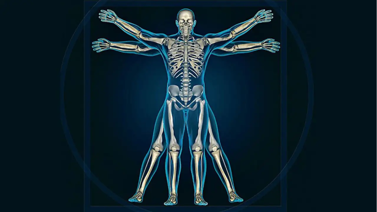
Roughly speaking, the human body is made up of
- A skeleton or bony frame-work.
- Appendages to the skeleton.
- Viscera or organs enclosed inside the cavities.
First then the skeleton or bony frame-work. This is what forms the frame-work for the support of softer structures (skin, muscles, vessels, nerves, etc.) and protection of the organs within the cavities of the body. Bones provide attachments for support of muscles and their tendons, ligaments, fibrous membranes, etc. They articulate with each other to form joints and are the levers in the movements at those joints.
The articulating surfaces of bones or those places which touch one another, are coated with a gristly material called articular cartilage. All bones are covered with fibrous vascular membrane called periosteum, whence blood vessels (nutrient arteries) enter into the bones through small foramen or openings on their surface. So long as this periosteum is healthy and intact, all branches in the bone can mend. If a piece of bone is medically sawn through, we will find that its surface is dense and hard (i.e., made up of compact tissue) and that its interior contains numerous spaces like a sponge (and so it is called cancellous or spongy tissue). The spongy tissue is filled with red marrow. In the case of long bones, the center, instead of being wholly filled with spongy tissue, is hollow, in which case, the hollow is called medullary cavity. According to their shape and size, bones are called— flat, short, irregular or long. The long bones have two extremities (epiphyses) or ends and a central shaft.
The bones of the skeleton are articulated or joined to each other by three means, viz., first, by sutures— i.e., by toothed edges of opposing bones, as in the case of the parietal bones that form the crown of the head; secondly, by means of cartilages or gristles, which sometimes, as in the case of joints form articular cartilages; thirdly, by ligaments or touch binders. Joints are surrounded by a very tough shiny sheet, called articular capsule, which prevents bones slipping out of place.
If we now study the Skeleton as a whole, we find that it is disposed thus
- Spinal Column as the central pivot
- Supporting the Skull
- Helping to form two cavities i.e., Thorax Abdomen
- Resting on Pelvic Girdle
- Having Upper and LowerAppendages or Extremities
The Spinal or Vertebral column is made up of
- 7 — Cervical or neck bones.
- 12 — Dorsal or back bones.
- 5 — Lumbar or loin bones.
- 5 — Sacral— or hip-bones (fused into one).
- 3 — Coccygeal or tail bones (fused into one).
Each spinal bone or vertebra has a central ring called vertebral foramen, to afford passage and support to the spinal cord. The spinal column then is a bony cagement —it supports on its top; the skull and it embodies inside the vertebral foramen and the spinal cord. The spinal column admits of very slight movement anteroposteriorly and laterally. Owing to faulty posture in sitting or standing, vertebral body gets unduly contorted, thereby producing deformity as kyphosis or stooping forward and scoliosis or laterally curved.
The twelve dorsal vertebrae have, on each side, twelve ribs attached to them. The ribs are called Costae. Of the twelve ribs, the first seven are connected in the front and central part of the body to the Sternum or Breast Bone. The sternum is made of three parts, whereof the upper two are bony and the lowermost, the Ensiform or Xiphoid Cartilage remains soft and elastic till about the end of middle age. After the age of 40, it begins to be converted into bone and becomes hard. It is important to remember this simple anatomical fact, inasmuch as, later in life, when it becomes hard and therefore prominent and palpable, people look upon it as a disease of the liver. Returning to the ribs, we find that the first seven ribs are called true ribs, because they are joined to the sternum. Of the remaining five, three upper ones are connected by their cartilage to the cartilage of the seventh rib; the last two are called floating or false ribs having one extremity free.
We now pass on to the skull or bones of the head. There are in the skull)
- 1 – Frontal bone—situated in front (forehead).
- 2 – Parietal bones (one on either side). By union, they form the crown or top of the head.
- 1 – Occipital bone—that forms the back of the head.
- 2 – Temporal bones— (one on each side). In these, occur the ear-opening (externaluditory meatus). Inside each are three small bones (ossicles) of middle ear.
- 1 – Sphenoid bone—wedged in between the temporal bones.
- 1 – Spongy, delicate, Ethmoid bone—situated behind the eyes on the top of the nose.
Then we pass down the Face. This part of our anatomy comprises the following bones.
- 2 – Superior Maxillary bones-helping to form the cheeks. They contain the upper row of teeth.
- 1 – Inferior Maxillary bone or mandible. This maxillary bone contains the lower row of teeth.
By their union, the two superior maxillary or jaw bones help to form the hard palate and the floor of the nose. The superior maxilla has a hollow cavity inside each, known as Antrum of Highmore. In fact the inside of each nose communicates with this cavity. The superior maxilla forms externally the prominence of the cheeks.
If an individual suffers from catarrh (running) of the nose, it causes pain in the region of the cheek-prominence by lighting up inflammation internally.
- 2 – Lachrymal bones-situated in front part of the orbit.
- 2 – Malar bones (one on each side)-situated on the outer and upper part the face—causing the prominence above the cheeks.
- 2—Palatine bones (one on each side)- situated in the back part of the nose.
- 2—Inferior Terbinated bones-one in the inside of each nasal cavity.
A separated reference is necessary to what is called the orbit. This is a round cavity, one on each side of the head, into which fits an eye-globe. It is made up of seven bones- the frontal, ethmoid, sphenoid, lachrymal, superior maxillary, palatine and malar bones.
Before leaving the head and face, it is important to draw attention to a few important surgical and medical aspects regarding them. The first thing to note is that the two points where the parietal bones come in contact with the Frontal and Occipital bones are called fontanelles in infants. The anterior fontanelle is a triangular gap found in infants under one year, where as frontal bone joins the two parietals. The posterior fontanelle is a lozenge-shaped gap, occurring also in infants, but of less than six months old, at the point of junction of the occipital with the parietal bones. These gaps get filled up with bony tissues as the child’s age advances. The second point of importance to note is that any jar given to the lower jaw may be transmitted to the head. This is severe and sudden, may even cause fracture of the skull at its base. Inside the skull is contained the Brain. We shall speak of the Brain hereafter.
We next take up the Pelvis. This is a basin intended to contain the genito-urinary organs and to give support to the spinal column. It is made up of
- 2—Os Innominata—one on either side.
- 1—Sacrum-(formed by fusion of five pieces of sacral vertebrae).
- 1—Coccyx-(formed by 4 coccygeal vertebrae).
The pelvis has an upper opening, called the ‘brim’ and a lower opening, called the pelvic ‘outlet’ Anteriorly the pelvis is guarded by symphysis pubis; it is here that the external organs of generation are attached. That part of pelvic or innominate bone on which we sit, is called ischium.
The pelvis in the male differs from the pelvis in the female. The points of difference are noted in the table below:
| Male Pelvis | Female Pelvis |
| Bones are stronger | Bones are lighter. |
| Muscular impression marked | They are less prominent |
| Cavity is deep and narrow | Cavity is roomy. |
| The brim is comparatively narrow. | Brim is wide. |
| Sacrum is much curved | Sacrum is less curved. |
| Pubic arch is narrow | Pubic arch is wider |
| Upper Extremity | Lower Extremity |
| Shoulder Girdle-Comprising · Clavicle (in front) · Scapula (behind) · Humerus (arm) · Badius (fore-arm) · Ulna (fore-arm) | Pelvic Girdle-Comprising · Os innominatum (in front) · Sacrum (behind) · Femur (thigh) · Fibula (leg) · Tibis with patella (leg). |
We now pass on to a consideration of the Extremities, and we shall do so by putting side by side in parallel column the homologous parts in the Hand and Foot
Hand
- Carpal Bones—Eight in number. Arranged in two rows. Those of the proximate row from the radial to the ulnar side are—Navicular, Lunate, Triquetral and Pisiform. Those of distal row are— Greater Multangular. Lesser Multangular. Capitate and Homate bone.
- Metacarpal bones—5 in all Phalanges 3 to each finger, and 2 for thumb.
Foot
- Tarsal Bones—Seven in number. Talus (Astragalus), Calcaneus, Navicular, 1st, 2nd & 3rd Cuneiform and Cuboid.
- Metatarsal bones—5 in all: Phalanges 3 to each toe except the first which has 2.
We now consider the appendages to the skeleton, and the first item is Muscle.
Muscle or flesh is a collection of stringy substances endowed with the property of contraction. A ‘Muscle’ as we know it, is made up of several bundles or muscle fibers, each fiber being made up of several cells. Muscles are said to originate from one bony spot and are said to be inserted into another bony spot. The central thickened part of a muscle is called the belly of the muscle, its ends (i.e., the points of insertion and origin) being made up of tendons or very strong shiny fibers that do not possess elasticity. Into muscles terminate the terminal divisions of nerves (nerve endings). Muscles as a rule, are richly supplied with blood-vessels. All muscles are on a condition of being in taut; this is known as their tonicity or state of tonic contraction. A muscle possesses tonus, elasticity, contractility and irritability.
Nerves are, like electric telegraph wires, vivid with life. They are shiny, hard, cord-like structures. They all originate either from the brain or the spinal cord and terminate on or are said to supply (be disturbed on) the skin or muscles or organs. They serve one of these three functions: (a) Those that are Motor nerves, send impulse to contract the muscles or groups of muscles in which they terminate, (b) Those that are Sensory, convey from the skin or viscera or wherever they happen to lie, to the brain sensations of pain or pleasure, heat or cold, touch or taste, as the case may be, and (c) Those nerves that are Trophic, carry nutritional impulses to the parts they supply, that is to say, if such a nerve is cut the parts supplied by it, suffer from want of nutrition; or in other words, those parts waste away.
Blood-vessels are divided into three classes, viz.
Arteries or those principal trunk-vessels that carry purified blood from heart to the distal parts of the body. These are endowed with muscular coat and as such can contract when cut or tom. They pulsate or carry on the wave of impulse imparted to the column of blood contained inside them, by the forcible contraction of the heart. When an artery is cut, blood flows out from it in jets or spurts, and such blood is bright scarlet colored.
Veins are principal vessels carrying back from distal parts of the body all the impure blood to the right side of the heart. They are devoid of muscular coat and as such when cut or tom, they gape and dark blood flows out of them in a continuous stream.
Arteries when they become smaller are called arterioles; and veins when they become smaller are called venules.
Capillaries Arterioles, when they enter into any part of the body, become smaller and smaller till they become so small as to possess only one-celled wall, they are then called capillaries. Capillaries then are, on the one hand, the final minute ramifications of arteries, and on the other the primal minute ramifications that will run on into venules. Capillaries are the end of arterial and beginning of venous systems. In them there is no throbbing pulsations of arteries but blood flows through them in a stream controlled by chemical and physical composition of blood (tension, viscosity and osmosis). Capillaries are like the spill areas of rivers. They carry arterial or pure blood to each cell of the part and gather up from those areas the impure blood to be transmitted into the right side of the heart. Like the spill area of rivers, the sectional area of capillary circulational is many times larger than the sectional area of the blood vessels. Blood flows quickly and in jerks through arteries, quick and in less jerkly spuers through arteries, slowly and without any jerk whatever through capillaries, through venules and veins into the heart.
Lymphatics: Lymph flows throughout the body and from each area of the body into the heart. Lymph-flow occurs chiefly through minute capillaries called lymphatic ducts, into a receptacle called receptaculum chyli, situated on the mid-line of the back of the abdominal cavity just in front of the 1st and 2nd lumbar vertebrea, to be finally carried into the right heart by the vessel called thoracic duct, which opens into the left innominate vein at its formation. During their passage from tissue to the receptaculum chyli, lymph ducts pass through lymphatic glands situated almost all over the body and especially so near the joints and in the pelvis. Lymph ducts are a sort of overflow pipe or feeders to the general circulation and lymphatic glands are the seats where any foreign or poisonous body circulating in lymph is combed out and arrested and disposed off.
Skin is the external covering of the body. Hairs and nails (in man) and scales or horn (of animals) are modification of skin. The skin is made up roughly of two layers: one — a layer of vascular connective tissue called the corium or cutis vera and the other—an external covering of epithelium called the epidermis or cuticle. The epidermis consists of 5 layers, the 3rd layer from within outwards is called the stratum ganulosum which contains the pigment and imparts colour to the skin. The cutis vera or true skin or dermis is abundantly supplied with nerves and blood-vessels and with fat and sweat glands. The sweat glands look like a coil of tubes and they possess a duct or pipe that opens on the surface of the skin by means of holes called pores. The fat, sebaceous or oil glands lie in the dermis and they too have a duct opening on the epidermis passing between the papillae; the ducts of sebaceous glands however open into the follicle of a hair. They secrete an oily fluid that helps to keep our skin smooth.
Hairs are composed of fish-scale like cells, enclosing central pith. They are sunk into the true skin, enclosed in a kind of bag (the hair follicle) and mounted on small ridges called papillae.
Nails represent the scaly superficial layer of skin in a condensed form. The bed on which the nail rests is the dermis layer of skin modified into very vascular parallel ridges.
- <a href="http://www.onlyhomeopathy.com/nervous-system-anatomy/”>NERVOUS SYSTEM
- <a href="http://www.onlyhomeopathy.com/circulatory-system-anatomy/”>CIRCULATORY SYSTEM
- <a href="http://www.onlyhomeopathy.com/digestive-system-anatomy/”>DIGESTIVE-SYSTEM
- <a href="http://www.onlyhomeopathy.com/respiratory-system-anatomy/”>RESPIRATORY SYSTEM
- <a href="http://www.onlyhomeopathy.com/urinary-system-anatomy/”>URINARY SYSTEM
- <a href="http://www.onlyhomeopathy.com/genital-system-anatomy/”>GENITAL SYSTEM
- THE EYES
- THE EARS
Comments are closed.